
植物研究 ›› 2026, Vol. 46 ›› Issue (4): 764-776.doi: 10.7525/j.issn.1673-5102.2026.04.016
• 研究论文 • 上一篇
李文珺, 罗莎, 史倩倩( )
)
收稿日期:2026-02-01
出版日期:2026-07-20
发布日期:2026-07-22
通讯作者:
史倩倩
E-mail:shiqianqian2005@163.com
作者简介:李文珺(2005—),女,本科生,主要从事紫斑牡丹色斑研究。
基金资助:
Wenjun LI, Sha LUO, Qianqian SHI()
Received:2026-02-01
Online:2026-07-20
Published:2026-07-22
Contact:
Qianqian SHI
E-mail:shiqianqian2005@163.com
摘要:
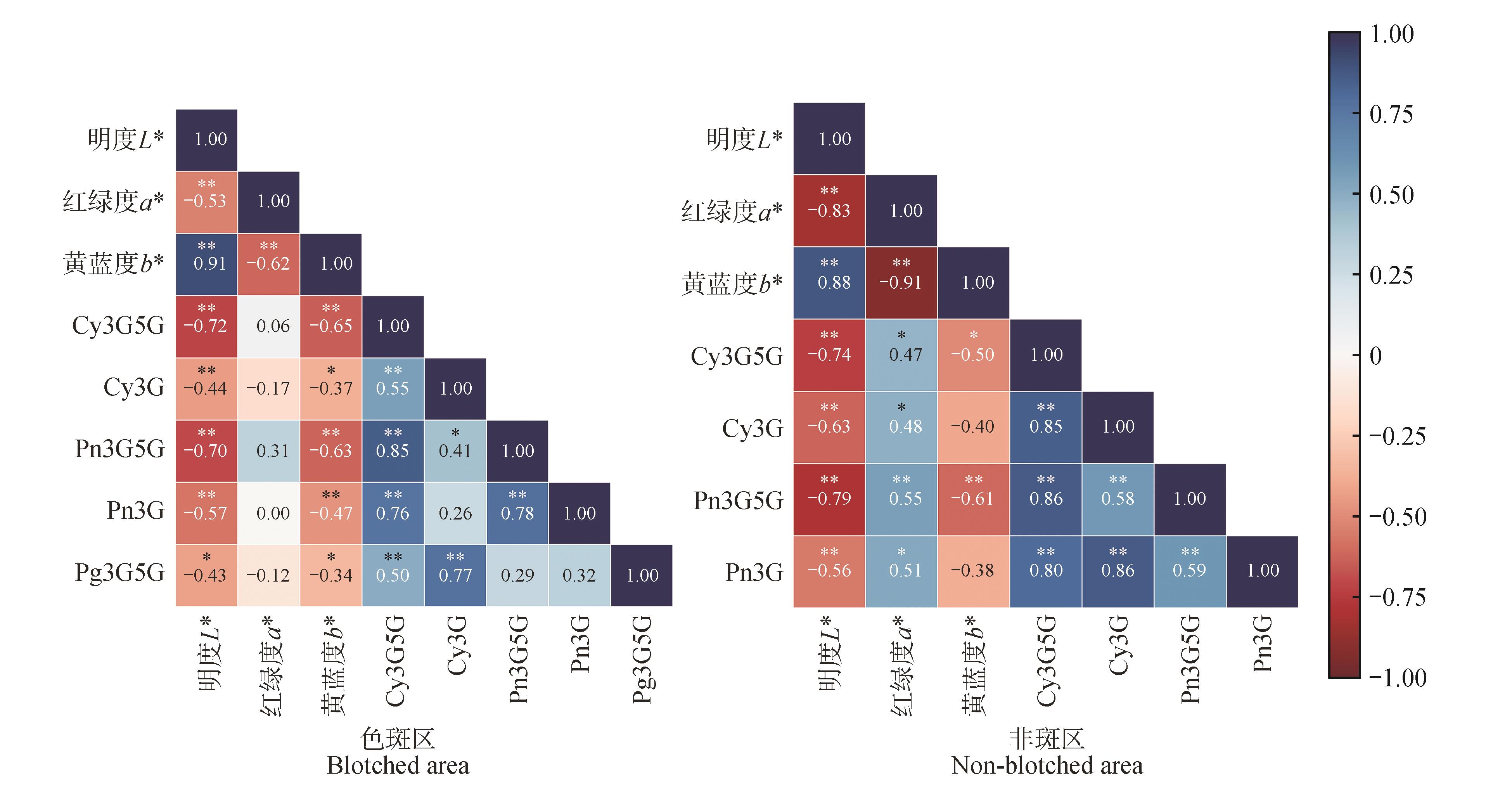
紫斑牡丹(Paeonia rockii)花瓣基部色斑是我国牡丹品种色斑性状的主要遗传来源。为明确色斑的时序发生、扩展模式及其与色素积累的关系,本研究系统观察了10个紫斑牡丹品种花瓣基部色斑在7个时期的形成情况。结果显示:S2时期花瓣基部出现单个色素细胞,并逐步形成以深色中心细胞为核心、浅色外围细胞环绕的色素细胞团。色斑以色素细胞团为基本结构单位进行扩展,其轮廓于S3时期初步形成,早于花瓣花色显现,且S6~S7时期仍存在色素细胞分裂高峰。S2~S3时期,色斑区L*(明度)和b*(黄蓝度)显著下降,a*(红绿度)显著升高。S2时期色斑区即可检测到花青苷,其中矢车菊素-3,5-O-二葡萄糖苷(Cy3G5G)和芍药花素-3,5-O-二葡萄糖苷(Pn3G5G)含量最高。线性回归分析表明,Pn3G5G含量对色斑L*、a*、b*变化具有较强解释力,是形成紫红色色斑的关键花青苷;天竺葵素-3,5-O-二葡萄糖苷(Pg3G5G)可能参与黑色色斑的形成。综上,紫斑牡丹色斑的形成是以色素细胞团扩展为结构基础、花青苷时空特异性积累为物质基础的动态发育过程。
中图分类号:
李文珺, 罗莎, 史倩倩. 紫斑牡丹色斑形成过程中表皮细胞变化及花青苷积累规律[J]. 植物研究, 2026, 46(4): 764-776.
Wenjun LI, Sha LUO, Qianqian SHI. Epidermal Cell Changes and Anthocyanin Accumulation Patterns during Paeonia rockii Blotch Formation[J]. Bulletin of Botanical Research, 2026, 46(4): 764-776.
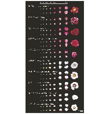
图1
10个紫斑牡丹品种S1~S7时期花瓣
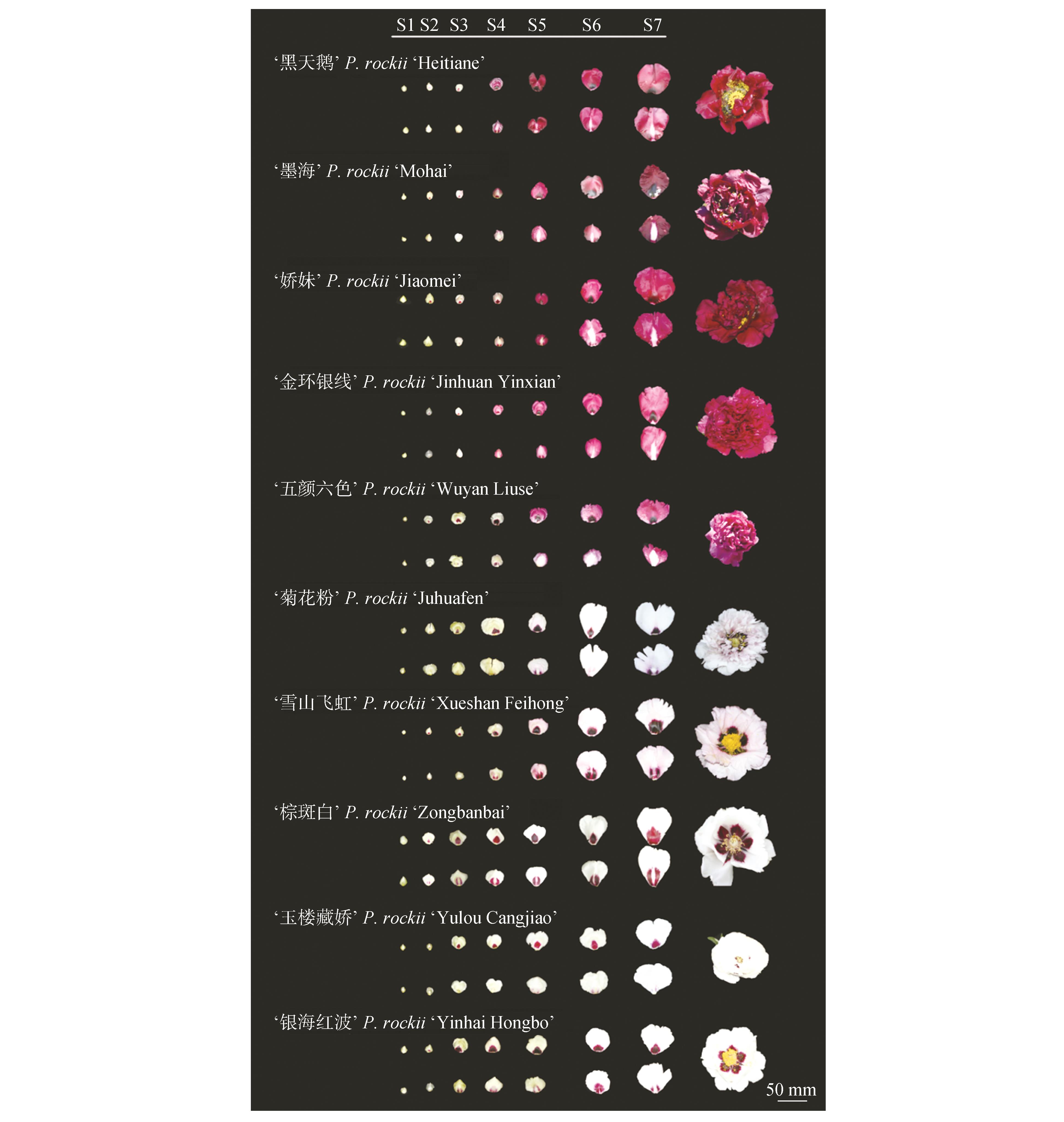
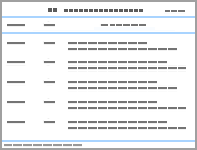
表1
紫斑牡丹花瓣色斑形成过程的时期划分
时期 Stage | 描述 Description | 对应牡丹物候期 Phenological period |
|---|---|---|
| S1 | 无色斑 | 立蕾期、小风铃前期 |
| S2 | 色斑逐渐显露,未形成完整轮廓 | 小风铃期、大风铃期 |
| S3 | 色斑轮廓初步形成,花色初显或将显未显 | 大风铃后期、圆桃前期 |
| S4 | 色斑增大加深,花色逐渐显露 | 圆桃期 |
| S5 | 色斑继续扩增,花色基本形成 | 圆桃后期、平桃期 |
| S6 | 色斑、花色已形成,面积仍有变化 | 破绽透色期、初开期 |
| S7 | 花开放 | 盛开期 |
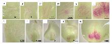
图2
S2~S3时期‘银海红波’花瓣色斑形成过程A~F.‘银海红波’花瓣的斑点扩散过程;G~K.‘银海红波’花瓣的左右斑点不对称扩散过程。
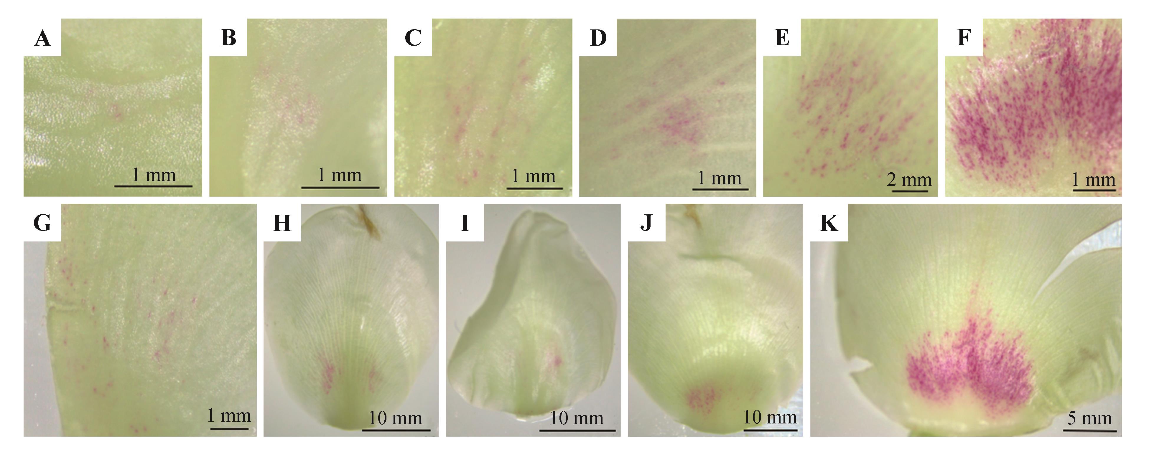
图3
S6时期紫斑牡丹品种花瓣基部色斑横纵切图A.‘银海红波’色斑区横切面;B.‘菊花粉’色斑区纵切面;C.‘五颜六色’色斑区横切面;D.‘玉楼藏娇’色斑区纵切面;E.‘菊花粉’色斑区色素细胞;F.‘菊花粉’色斑区色素细胞;G.‘雪山飞虹’色斑区色素细胞。
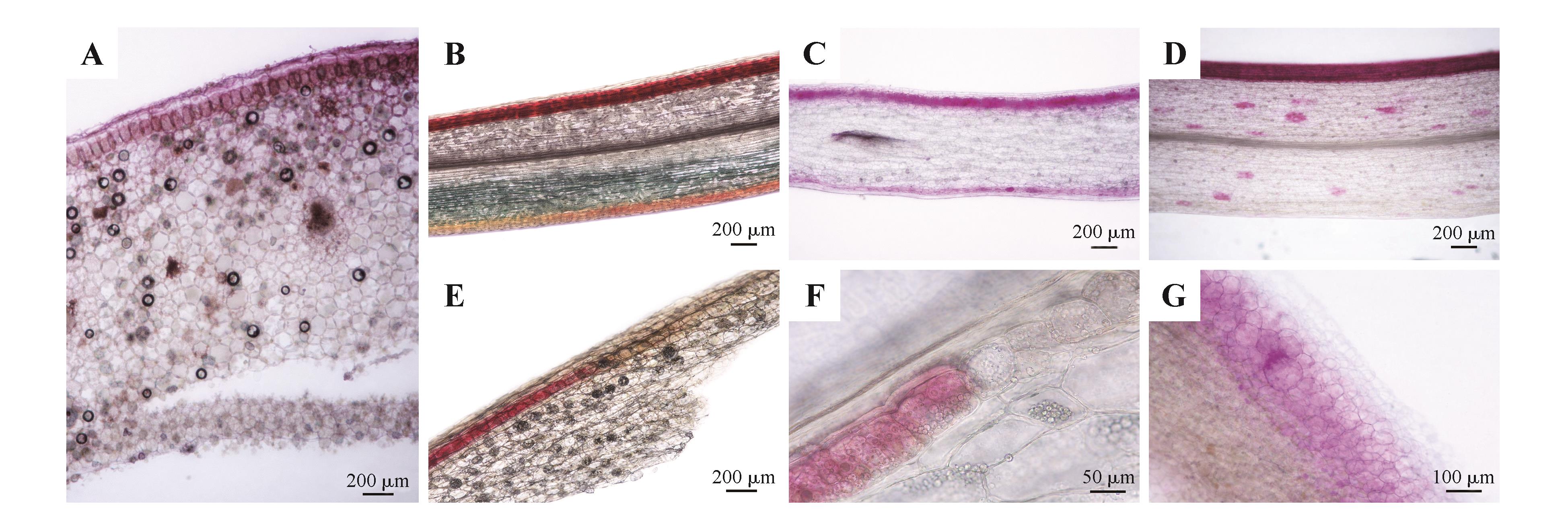
图4
S1~S7时期10个紫斑牡丹品种色斑区上表皮解剖结构
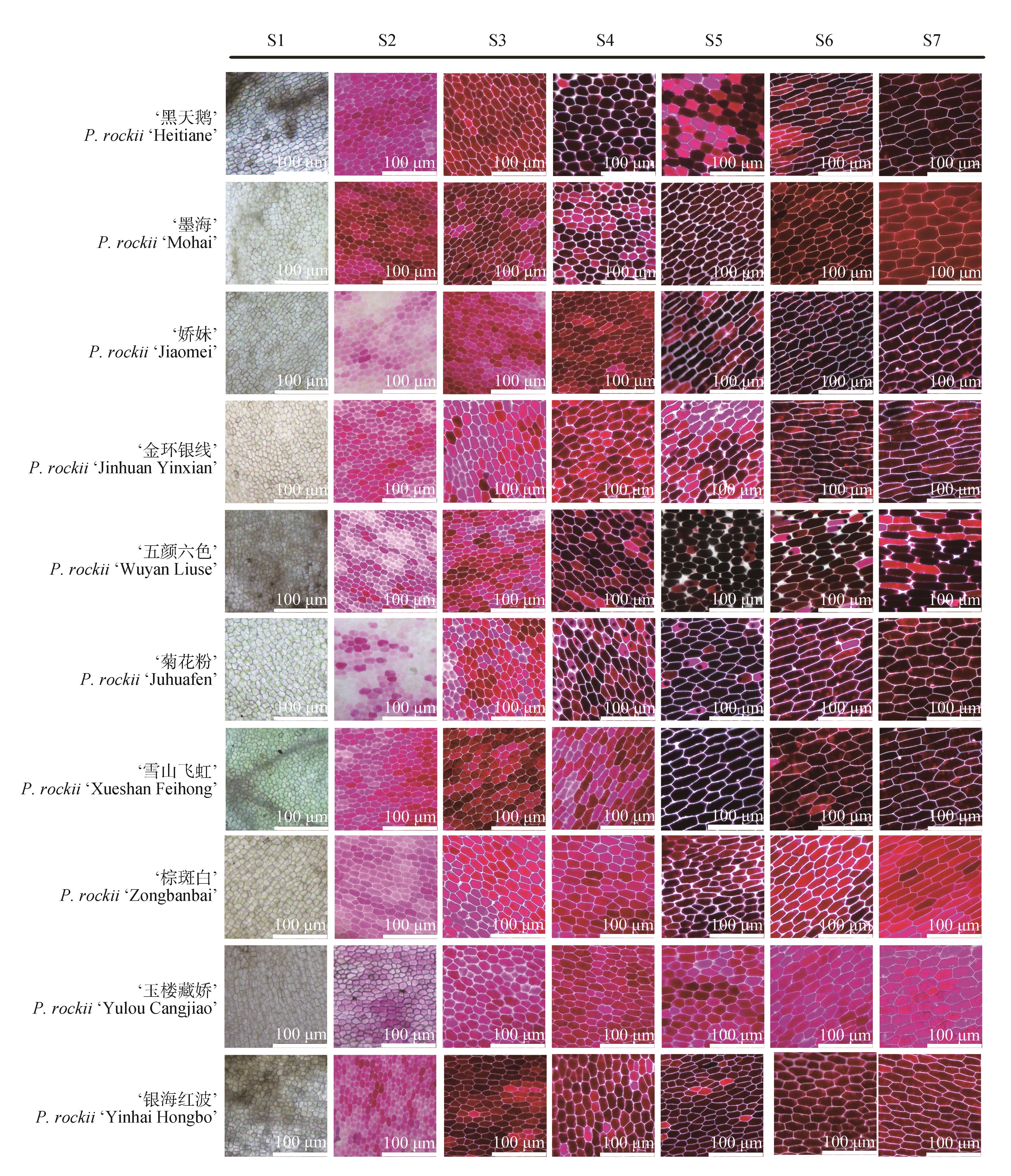
图5
紫斑牡丹中心色素细胞及色素细胞团A.‘银海红波’色素细胞; B.‘菊花粉’色素细胞; C.‘银海红波’色素细胞; D.‘五颜六色’色斑基部色素细胞;E.‘棕斑白’色斑顶部色素细胞; F.‘墨海’色斑基部色素细胞; G.‘雪山飞虹’色斑顶部色素细胞。A~C.S2前期; D、E.S2后期;F、G.S6期。图中标注:1.单个色素细胞;2.未成型色素细胞团;3.色素细胞团;4.色素细胞团连接成片。
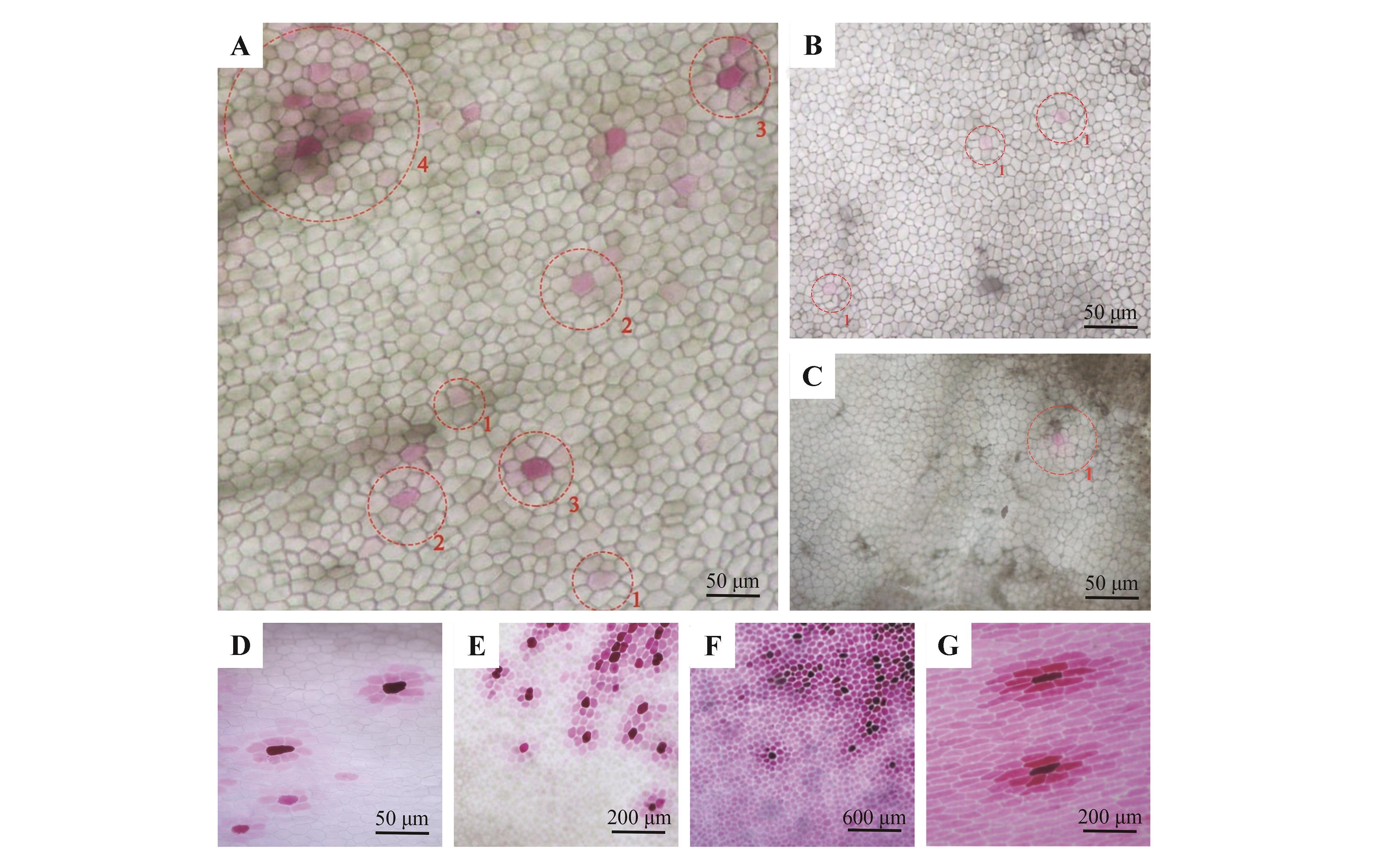
图6
S6时期紫斑牡丹品种色斑基部到顶部上表皮拼接图A.‘五颜六色’; B.‘银海红波’; C.‘棕斑白’。左端为色斑基部,右端为色斑顶部。
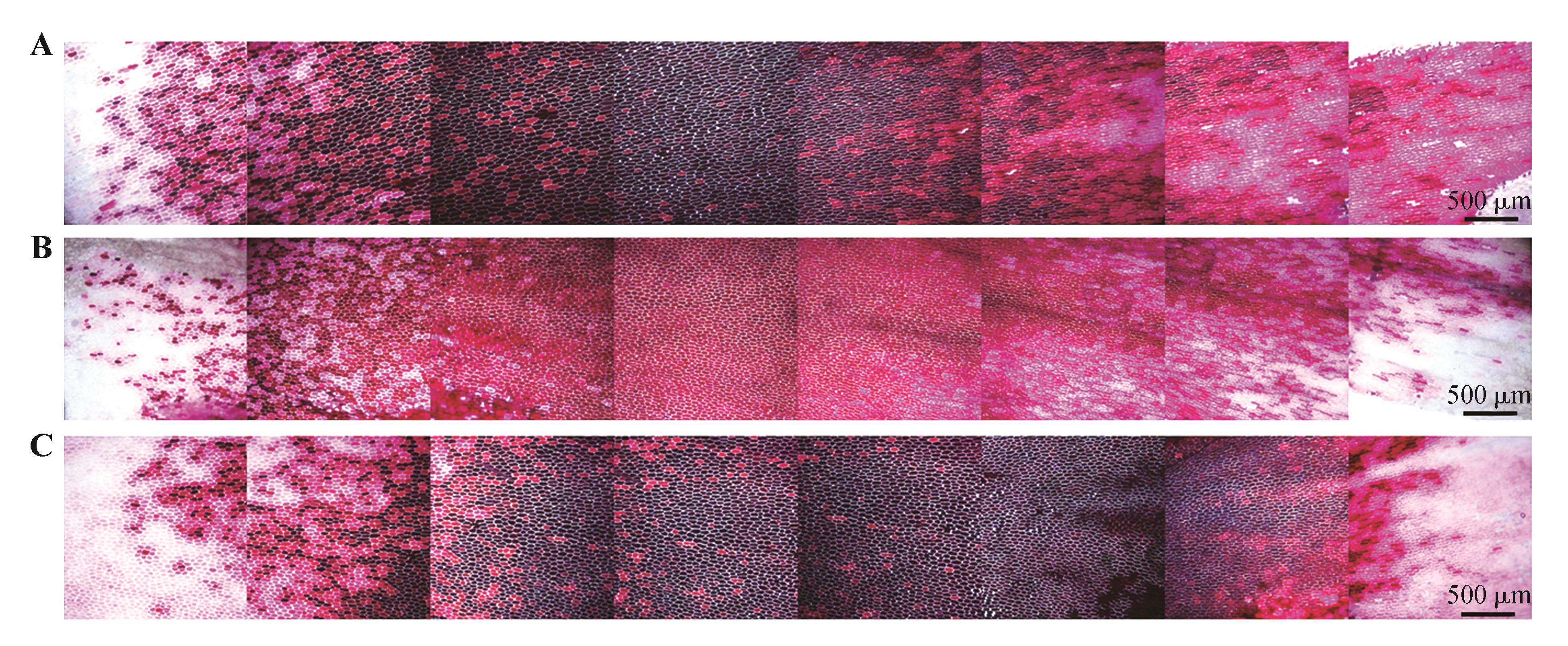
图7
紫斑牡丹品种在S1~S7时期的色斑细胞数目、面积和斑瓣比的变化
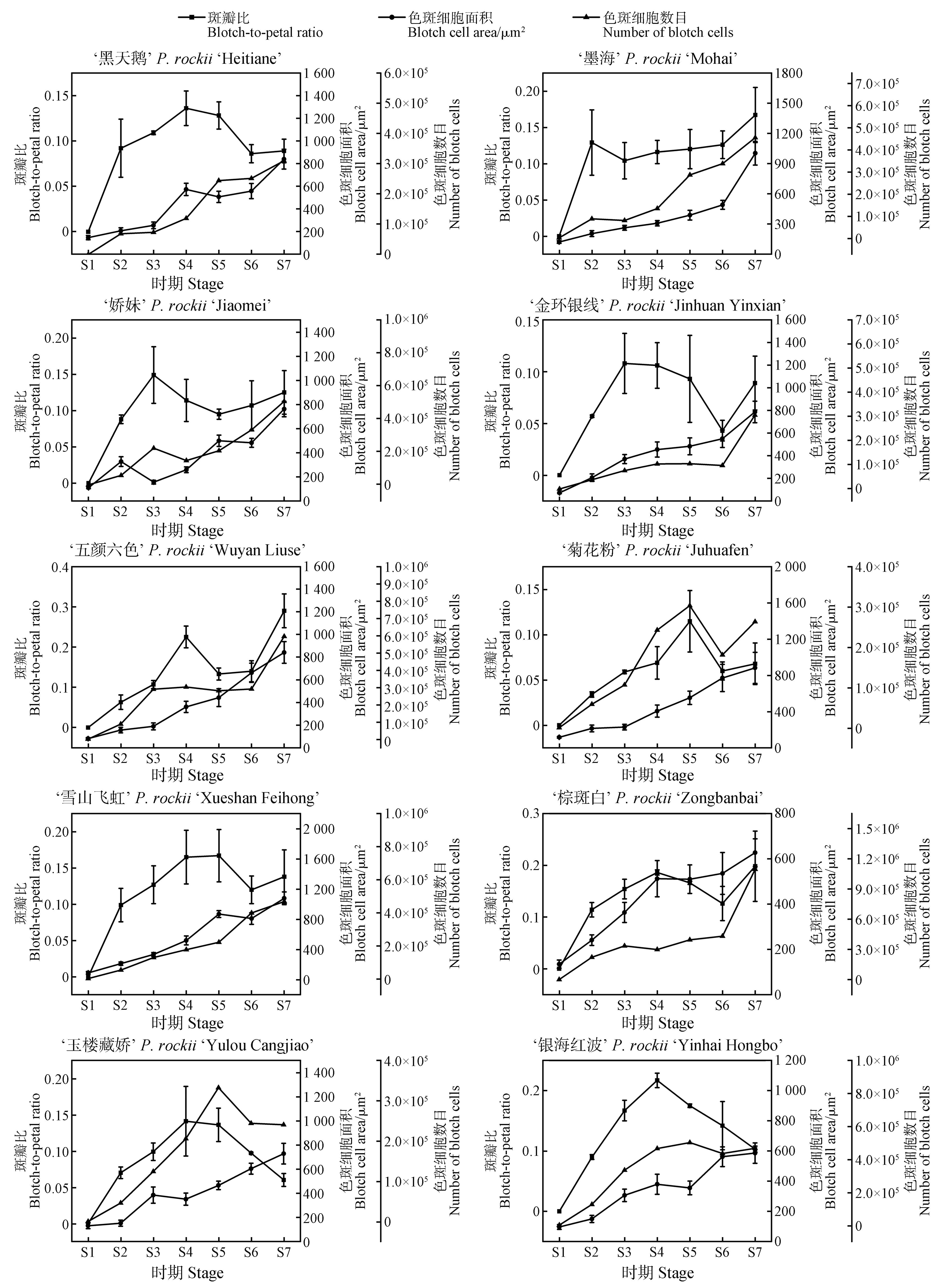
图8
紫斑牡丹品种色斑形成过程中的花色值箱线图箱内横线表示中位数,方形标记表示均值。
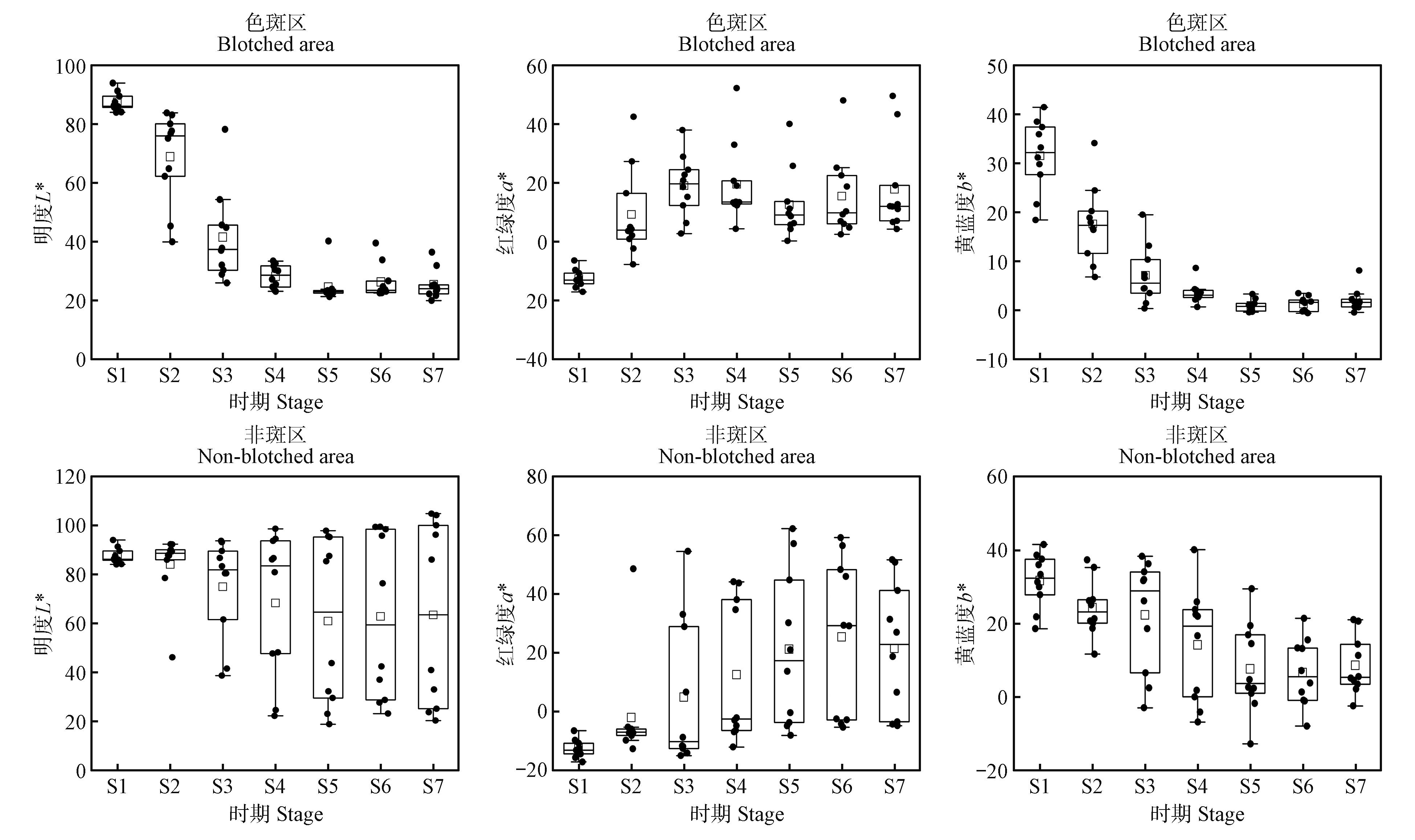
图9
紫斑牡丹品种花瓣色斑形成过程中色斑区与非斑区花青苷质量分数B.色斑区;N.非斑区。
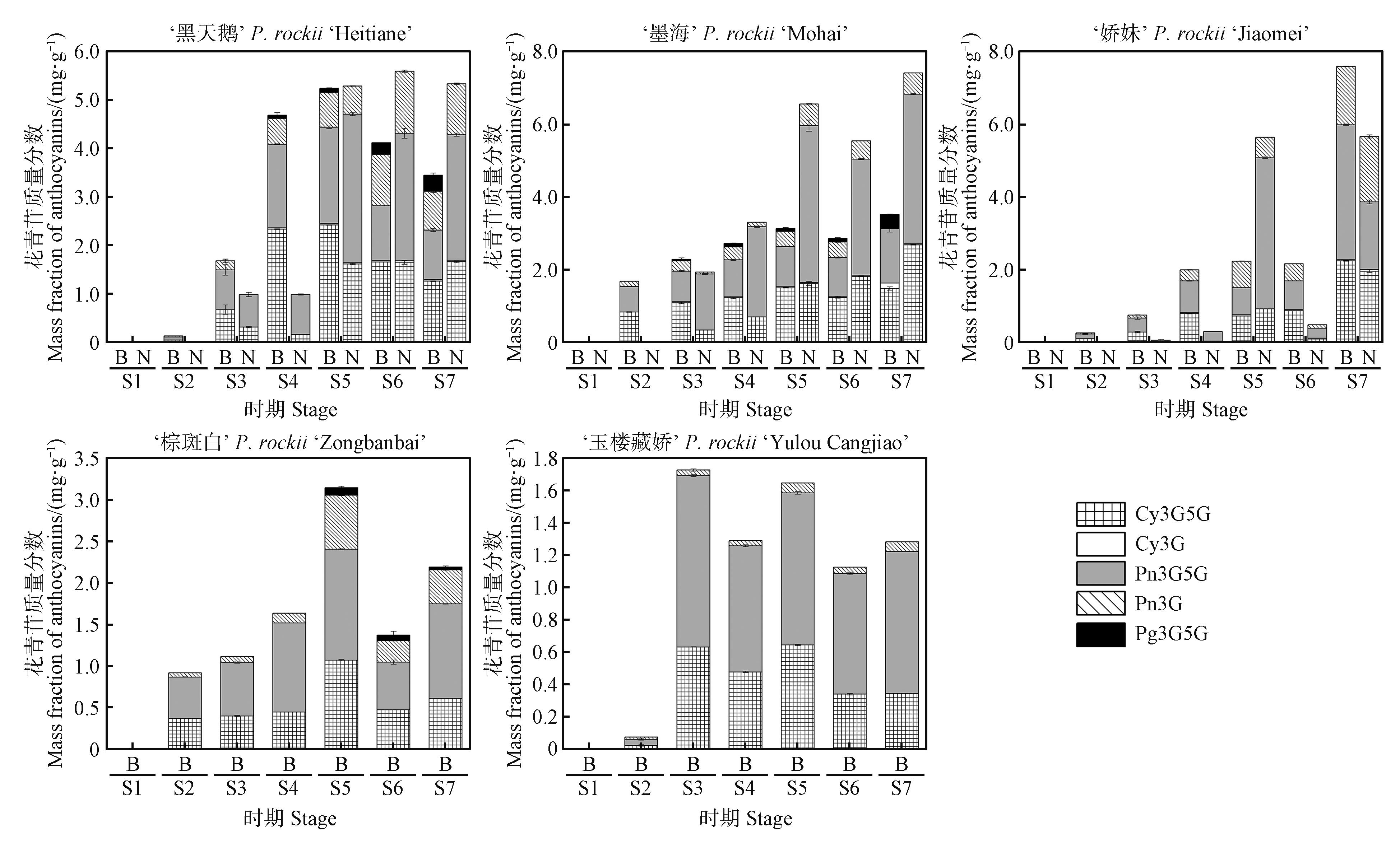
图10
紫斑牡丹品种花瓣色斑区和非斑区的花色值与花青苷含量的相关性分析*. P<0.05;**. P<0.01.
| [1] | 程金水.园林植物遗传育种学[M].北京:中国林业出版社,2000:81-82. |
| Cheng Jinshui.Genetics and breeding of garden plants[M].Beijing:China Forestry Publishing House,2000:81-82. | |
| [2] | Fairnie A L M, Yeo M T S, Gatti S,et al.Eco-Evo-Devo of petal pigmentation patterning[J].Essays in Biochemistry,2022,66(6):753-768. |
| [3] | Zhang Y Z, Cheng Y W, Ya H Y,et al.Transcriptome sequencing of purple petal spot region in tree peony reveals differentially expressed anthocyanin structural genes[J].Frontiers in Plant Science,2015,6:964. |
| [4] | 成仿云.中国紫斑牡丹[M].北京:中国林业出版社,2005:88. |
| Cheng Fangyun.Chinese flare Mudan[M].Beijing:China Forestry Publishing House,2005:88. | |
| [5] | Wang L S, Hashimoto F, Shiraishi A,et al.Phenetics in tree peony species from China by flower pigment cluster analysis[J].Journal of Plant Research,2001,114(3):213-221. |
| [6] | Wang L S, Hashimoto F, Shiraishi A,et al.Chemical taxonomy of the Xibei tree peony from China by floral pigmentation[J].Journal of Plant Research,2004,117(1):47-55. |
| [7] | Zhang J J, Wang L S, Shu Q Y,et al.Comparison of anthocyanins in non-blotches and blotches of the petals of Xibei tree peony[J].Scientia Horticulturae,2007,114(2):104-111. |
| [8] | Shi Q Q, Yuan M, Wang S,et al.PrMYB5 activates anthocyanin biosynthetic PrDFR to promote the distinct pigmentation pattern in the petal of Paeonia rockii [J].Frontiers in Plant Science,2022,13:955590. |
| [9] | Shi Q Q, Li L, Zhang X X,et al.Biochemical and comparative transcriptomic analyses identify candidate genes related to variegation formation in Paeonia rockii [J].Molecules,2017,22(8):1364. |
| [10] | Qi Y Y, Lou Q, Li H B,et al.Anatomical and biochemical studies of bicolored flower development in Muscari latifolium [J].Protoplasma,2013,250(6):1273-1281. |
| [11] | Schindelin J, Arganda-Carreras I, Frise E,et al.Fiji:an open-source platform for biological-image analysis[J].Nature Methods,2012,9(7):676-682. |
| [12] | Zhang J, Wang L S, Gao J M,et al.Determination of anthocyanins and exploration of relationship between their composition and petal coloration in crape myrtle (Lagerstroemia hybrid)[J].Journal of Integrative Plant Biology,2008,50(5):581-588. |
| [13] | Hanbury A, Serra J.Mathematical morphology in the cielab space[J].Image Analysis & Stereology,2002,21(3):201. |
| [14] | Luo X N, Sun D Y, Wang S,et al.Integrating full-length transcriptomics and metabolomics reveals the regulatory mechanisms underlying yellow pigmentation in tree peony (Paeonia suffruticosa Andr.) flowers[J].Horticulture Research,2021,8:235. |
| [15] | Suzuki K, Suzuki T, Nakatsuka T,et al.RNA-seq-based evaluation of bicolor tepal pigmentation in Asiatic hybrid lilies (Lilium spp.)[J].BMC Genomics,2016,17(1):611. |
| [16] | Wang Z, Li X, Chen M M,et al.Molecular and metabolic insights into anthocyanin biosynthesis for spot formation on Lilium leichtlinii var.maximowiczii flower petals[J].International Journal of Molecular Sciences,2023,24(3):1844. |
| [17] | Xu L F, Yang P P, Feng Y Y,et al.Spatiotemporal transcriptome analysis provides insights into bicolor tepal development in Lilium “tiny padhye”[J].Frontiers in Plant Science,2017,8:398. |
| [18] | Zhu J, Wang Y Z, Wang Q Y,et al.The combination of DNA methylation and positive regulation of anthocyanin biosynthesis by MYB and bHLH transcription factors contributes to the petal blotch formation in Xibei tree peony[J].Horticulture Research,2023,10(7):uhad100. |
| [19] | Wilts B D, Rudall P J, Moyroud E,et al.Ultrastructure and optics of the prism-like petal epidermal cells of Eschscholzia californica (California poppy)[J].New Phytologist,2018,219(3):1124-1133. |
| [20] | 林登贵,曾丽,裴峰,等.万寿菊营养器官及舌状花解剖结构的研究[J].上海交通大学学报(农业科学版),2015,33(4):59-64. |
| Lin Denggui, Zeng Li, Pei Feng,et al.Anatomical structures of vegetative organs and ray floret in Tagetes erecta L.[J].Journal of Shanghai Jiao Tong University(Agricultural Science),2015,33(4):59-64. | |
| [21] | 殷涵泰,尹俊梅,廖易,等.基于秋石斛花朵颜色、色素分布及表皮细胞形态的表型分类[J].园艺学报,2021,48(10):1907-1920. |
| Yin Hantai, Yin Junmei, Liao Yi,et al.Phenotype classification based on flower color,pigment distribution and epidermal cell shape of Dendrobium hybrids[J].Acta Horticulturae Sinica,2021,48(10):1907-1920. | |
| [22] | Luan Y T, Tang Y H, Wang X,et al.Tree peony R2R3-MYB transcription factor PsMYB30 promotes petal blotch formation by activating the transcription of the anthocyanin synthase gene[J].Plant and Cell Physiology,2022,63(8):1101-1116. |
| [23] | Kicheva A, Briscoe J.Developmental pattern formation in phases[J].Trends in Cell Biology,2015,25(10):579-591. |
| [24] | Davies K M, Albert N W, Schwinn K E.From landing lights to mimicry:the molecular regulation of flower colouration and mechanisms for pigmentation patterning[J].Functional Plant Biology,2012,39(8):619-638. |
| [25] | Li Y, Kong F, Liu Z A,et al.PhUGT78A22, a novel glycosyltransferase in Paeonia ‘He Xie’, can catalyze the transfer of glucose to glucosylated anthocyanins during petal blotch formation[J].BMC Plant Biology,2022,22:405. |
| [1] | 胡昕怡, 许黎明, 覃伯韬, 安倚东, 蒋国凤. 睡莲科植物叶与陆生草本植物叶供耗水特征差异[J]. 植物研究, 2025, 45(5): 675-685. |
| [2] | 唐楠, 李苗苗, 唐道城. 青藏高原不同海拔高度下全缘叶绿绒蒿叶表皮特征研究[J]. 植物研究, 2019, 39(2): 161-168. |
| [3] | 徐莉, 成仿云, 钟原. 牡丹胚培养快速成苗技术研究[J]. 植物研究, 2017, 37(5): 690-699. |
| [4] | 王崑仑1;赵修华1;祖元刚1*;李家磊2. 白藜芦醇在几种植物中各部位的分布[J]. 植物研究, 2015, 35(4): 638-640. |
| [5] | 张衷华1,2;唐中华1;杨逢建1*;王化3;张军1;祖元刚1. 两种主要油用牡丹光合特性及其微环境影响因子分析[J]. 植物研究, 2014, 34(6): 770-775. |
| [6] | 梁文斌;赵丽娟;李家湘*;肖健. 湖南安息香属植物的叶片比较解剖学研究[J]. 植物研究, 2014, 34(2): 148-158. |
| [7] | 张曼曼;努尔买买提·莫明;王虹*;布佐日古丽·喀迪尔;姜彦成. 委陵菜属锥状花柱组植物叶表皮微形态特征的研究[J]. 植物研究, 2013, 33(1): 7-17. |
| [8] | 朱艳;徐增莱*;汪琼;王贞;史云云. 不同倍性盾叶薯蓣(Dioscorea zingiberensis)叶表皮形态的比较[J]. 植物研究, 2010, 30(3): 267-272. |
| [9] | 许玉凤;张柯;王文和;李楠. 鸢尾属(Iris)植物叶片表皮微形态特征的研究[J]. 植物研究, 2008, 28(5): 547-551. |
| [10] | 洪涛, 张家勋, 李嘉珏, 赵文忠, 李明瑞. 中国野生牡丹研究(一)芍药属牡丹组新分类群[J]. 植物研究, 1992, 12(3): 223-234. |
| 阅读次数 | ||||||
|
全文 |
|
|||||
|
摘要 |
|
|||||
